 Ground Floor
Suite 3-6, 100 Victoria Parade
East Melbourne VIC 3002
Ph: 03 9667 1667
Fax: 03 9667 1666
Ground Floor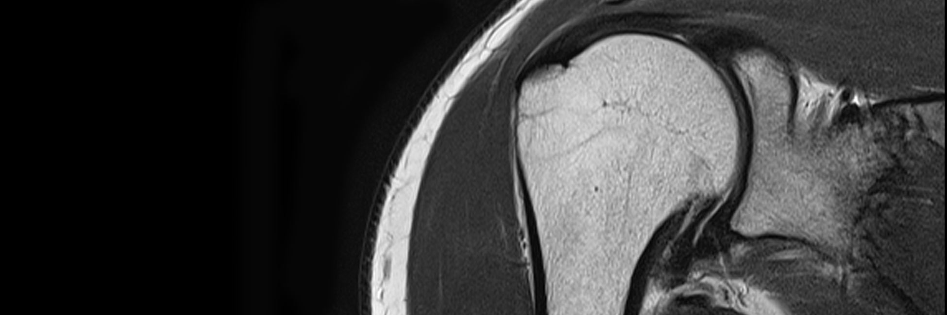


Magnetic Resonance Imaging (MRI) is the most sophisticated imaging modality that has revolutionised modern radiology and overall is considered to be the best type of radiological scan.
The human body is made up of approximately 70% water and MRI takes advantage of these hydrogen atoms in the body to create extremely high quality imaging of body tissue.
MRI Scanners use a large and powerful magnet to align water molecules in the body in a particular direction. Radiofrequency (energy) waves, similar to those used in a commercial radio, enter the body, manipulating the magnetic alignment of the molecules. When these radiofrequency waves are switched off, the water molecules then release the energy that was imparted to them, a process known as “resonance”.
The manner in which molecules release this energy differs on the specific type of tissue and is dependent on other factors, including the local magnetic field in our body at the molecular level. This released energy is then detected by a coil as a signal, which is then digitised and processed to form an image.
MRI technology uses energy in the radiowave spectrum and thus does not result in any radiation exposure. For this reason, MRI is of particular use in the evaluation of paediatric patients, whose organs are more radiosensitive than adults.
MRI scanners use powerful magnets and radio waves which aligns hydrogen atoms in the body in one direction.
Radiation exposure equivalent to:
Days of Natural
Background Radiation
vs
Days of Flying
*ultra-low dose CT scanner
(Siemens SOMATOM go.Up)


Versatile and Sensitive.
Magnetic Resonance Imaging is extremely versatile and sensitive, with the ability to differentiate between different body tissues, such as the grey and white matter of the brain, cartilage, bone, tendons, fluid and fat.
This is one of the advantages of MRI over ultrasound. For example, an ultrasound scan will provide excellent resolution of the rotator cuff tendons of the shoulder, but is unable to exclude any problem inside the shoulder joint, such as a cartilage or labral tear, arthritis or bone injury. MRI can look at all these structures.
Bowel MRI
The applications of MRI are ever expanding, and now may be even used to assess the small and large bowel for masses and obstruction, as well as conditions such as Crohn’s disease and cancer.


dGEMRIC MRI.
MRI has the ability to even identify early cartilage breakdown at the molecular level prior to the development of a visible defect and overt arthritis utilising a technique known as delayed Gadolinium Enhanced Magnetic resonance Imaging of Cartilage, or dGEMRIC.
Similarly, though CT can obtain excellent views of the brain, MRI can differentiate with far greater resolution the grey and white matter of the brain.
Further Information.
Referring doctors are welcome to discuss with our radiologists the imaging needs of their patients and whether MRI is suitable for their patient’s medical condition.